Hearing a long medical term before surgery can be unsettling. If your surgeon mentioned an “intra-operative cholangiogram,” you may be wondering whether something is wrong. In most cases, it is simply a way of looking at the bile ducts during an operation so that your surgeon can proceed with more information. Understanding it ahead of time can make the day of surgery feel far less intimidating.
What it is
A cholangiogram is an X-ray study of the bile ducts, the small tubes that carry bile from the liver and gallbladder into the small intestine. “Intra-operative” means it is performed during surgery, most often during laparoscopic gallbladder removal (cholecystectomy). A contrast dye is gently introduced into the bile duct system, and real-time X-ray images show the outline of the ducts on a monitor.
Why it may be done
The bile ducts are small, and their layout varies from person to person. The images can help a surgeon in several ways:
- Confirm the anatomy of the ducts before they are cut or clipped.
- Check for stones that may have moved out of the gallbladder into the common bile duct.
- Identify a leak or injury to a duct.
- Clarify the picture when inflammation makes the anatomy hard to see.
Who it affects
People having gallbladder surgery for gallstones, gallbladder inflammation, or related problems may be candidates. It is not performed for every patient. Some surgeons use it routinely, while others use it selectively, for example when blood tests suggest a stone in the duct, when a patient has had pancreatitis or jaundice, or when the anatomy is unclear during surgery.
How the procedure works
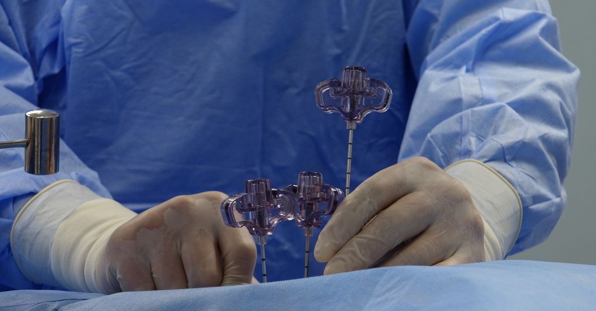
While you are under general anesthesia, the surgeon places a very thin catheter into the cystic duct, which connects the gallbladder to the main bile duct. Contrast dye flows through the catheter, and X-ray images are taken. If the ducts fill normally and the dye flows into the intestine, the surgeon can proceed. If a stone or blockage appears, the surgeon can decide how best to address it. You will not feel the test, and it usually adds only a short amount of time to the operation.
What the research suggests
Surgeons and professional societies continue to discuss when routine cholangiography is best. Studies have suggested that imaging can help recognize bile duct injuries earlier and may reduce their severity, while other reviews have found that selective use is reasonable for many patients. Because evidence and individual risk differ, the decision is usually personalized rather than automatic.
Risks and considerations
The test is generally considered safe. Possible concerns include a reaction to contrast dye, a small amount of added radiation exposure, and a slightly longer operating time. Rarely, placing the catheter can cause irritation of the duct. Your surgeon can explain how these apply to you.
Questions and tips before surgery
- Ask why a cholangiogram is being considered in your specific case.
- Tell your surgeon about any allergy to contrast dye or iodine.
- Mention any history of kidney problems, pregnancy, or prior biliary surgery.
- Ask what happens if the images show a stone or an unclear duct.
- Review recovery expectations and warning signs before you go home.
Surgery brings natural questions, and you deserve clear answers. If you have concerns about gallbladder problems or an upcoming procedure, the practice of Aldo Gamarra, MD in Lancaster, CA can talk with you about your situation. Call (661) 579-6088 or reach out through our contact page.